

Many people still view root canal treatment as one of the most unpleasant dental procedures. Fear of pain, lengthy treatment, and poor results leads people to put off visiting a dentist until the last minute. But today, root canal treatment is a precise, comfortable, and technologically advanced procedure, the main goal of which is to relieve pain and preserve natural teeth for many years.
More and more clinics are switching to microscopic treatment. A dentist simply can’t see the smallest root canal branches, hidden cracks, or remaining infection with the naked eye. Such details often lead to persistent or recurrent infection and subsequent tooth extraction. A microscope allows the dentist to literally “look” inside the tooth, perform treatment with maximum precision, and reduce the risk of complications.
In this article, we’ll discuss when microscopic root canal treatment is necessary, how the procedure is performed, why it’s considered more effective, and whether it can actually help save teeth even in difficult situations.
When is Root Canal Treatment under a Microscope Necessary?
Microscopic root canal treatment is necessary in several situations.
The most common cause is deep decay that has reached inside the tooth and affected the pulp. When bacteria penetrate the tooth, inflammation occurs, causing pain, swelling, and sensitivity.
Treatment is also required after an injury. If a tooth is cracked or broken, the pulp may become exposed, creating a risk of infection.
Sometimes, treatment is necessary after previous dental procedures. Poorly performed treatment can lead to persistent or recurrent infection and the need for retreatment of root canals.
In severe cases, the infection spreads around the root, causing a periapical lesion. Without timely intervention, the patient may require a tooth extraction.
What is Root Canal Treatment under a Microscope?
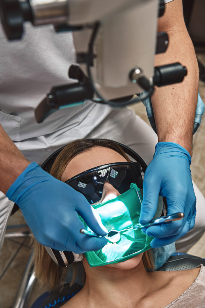
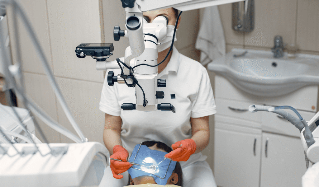
Root canal treatment under microscope is a modern technique in which the dentist uses a specialized endodontic microscope to magnify the working area.
This treatment is also called microscope assisted root canal treatment, as all stages of the procedure are performed under magnification.
The main purpose of the microscope is to allow the dentist to see the smallest details of the root system and perform treatment with maximum precision.
A microscope helps detect:
- microcracks;
- hidden infections;
- additional canals;
- calcified areas;
- instrument residue;
- poorly sealed canals.
Modern microscopes provide magnification from 6 to 25 times. This high magnification significantly improves the quality of treatment.
How Does Root Canal Treatment under a Microscope Work?
During endodontic treatment under a microscope, the dentist uses magnification and modern instruments to clean and fill canals. First, a diagnosis is made. Digital dental imaging and X-ray examinations are used to assess the condition of the tooth. The microscope allows the dentist to clearly visualize the internal structure of the tooth.
Precision root canal therapy is performed under high-quality anesthesia, so the patient feels virtually no pain. Furthermore, working under a microscope allows the dentist to work with greater precision, which may speed up recovery after the procedure.
What Does Treatment under a Microscope Look Like, Step by Step?
Many patients worry that root canal treatment is long, complicated, and painful. In practice, modern endodontics under a microscope is much more comfortable than commonly thought. Below, we’ll look at the procedure step by step.
Diagnosis and Planning
The first stage is diagnosis and endodontic treatment planning. The doctor evaluates the condition of the roots, tissues, and the presence of inflammation using an X-ray of the tooth.
At this stage, the following is determined:
- the shape of the roots;
- the number of canals;
- the presence of cracks;
- signs of infection.
Anesthesia and isolation of the treatment area
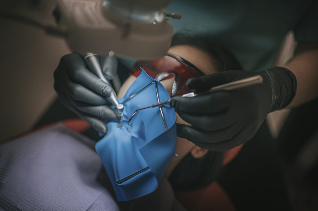
Next, anesthesia is administered, and the treatment area is prepared. A rubber dam is used to protect the tooth, helping to keep the treatment area clean during the procedure. After this, the dentist gains access to the pulp chamber.
Canal Cleaning and Shaping
This is one of the most important stages.
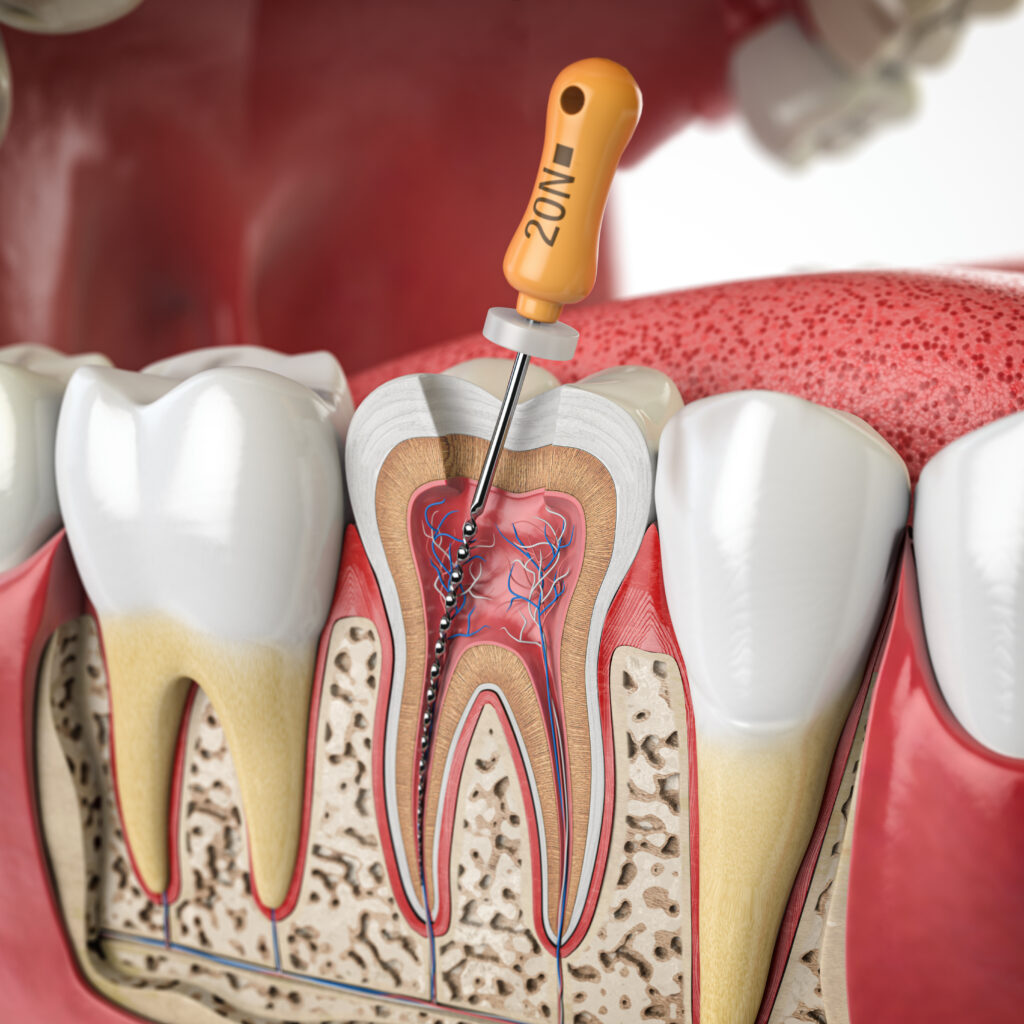
During canal treatment under the microscope, the doctor removes inflamed tissue and cleans the canals.
The microscope helps locate:
- hidden canals;
- calcified areas;
- cracks;
- additional canals.
Many teeth have complex anatomy and a complex root system that is difficult to see without magnification. The magnification provided by the microscope helps the doctor thoroughly clean the canals. At this stage, damaged tissue and pulp from the canals are removed, followed by disinfection.
The main focus is on:
- canal cleaning;
- removing bacteria;
- cleaning root canal contents;
- protecting tooth roots from bacteria.
Filling and Restoration
After cleaning, the dentist seals the canals. The main goal is to fill the root canals and prevent re-infection. After this, the tooth is reconstructed to restore function and aesthetics. A properly restored tooth after root canal treatment can last for many years.
Advantages of Treatment under a Microscope
Microscopic treatment is considered one of the most reliable and safest methods of modern endodontics. Let’s look at the main advantages of this technology for the patient.
- Greater treatment precision. Magnification allows the doctor to better view the treatment area.
- Magnification of the treatment helps to see details that are impossible to see with the naked eye.
- This treatment reduces the risk of complications because the doctor can detect hidden problems earlier.
- The treatment minimizes the risk of persistent or recurrent infection.
- Magnification improves cleaning accuracy and helps reduce the risk of missed canals and reinfection.
- A microscope helps to better treat the chamber and root canals.
- The doctor’s precise work often results in shorter treatment times and a more comfortable recovery.
Who is Suitable for Microscopic Treatment?
This treatment is recommended for patients with:
- severe pain;
- deep inflammation;
- complex anatomy;
- persistent or recurrent infection;
- cracked teeth.
Treatment is especially important for:
- curved roots;
- complex anatomy;
- pulp infection or irreversible pulpitis.
How Much Does Root Canal Treatment under a Microscope Cost?
The cost of treatment depends on several factors:
- the complexity of the case;
- the location of the tooth;
- the number of canals;
- the degree of inflammation;
- the need for retreatment.
Molars are generally more expensive due to their complex anatomy and the large number of canals. The price may also increase if retreatment or restoration of a severely damaged tooth is required.
Root canal retreatment at our clinic is performed exclusively using a microscope, as this approach allows for thorough root canal cleaning and reduces the risk of persistent or recurrent infection.
The exact cost is determined after diagnosis, as each clinical condition requires an individual approach.
Is it Possible to Repeat the Treatment under a Microscope?
Repeated infected root canal treatment is one of the main advantages of microscopic endodontics. In many cases, a tooth previously considered “hopeless” can now be successfully saved thanks to magnification.
With a microscope, the dentist can:
- remove old filling material;
- find missed or overly narrow canals;
- eliminate hidden foci of infection;
- correct errors in previous treatment;
- carefully treat complex areas of the root system.
Why Doesn’t Every Dentist Use a Microscope?
The main reasons why not every dentist works with a microscope are:
- high cost of equipment;
- need for additional training;
- difficulty working under magnification;
- time-consuming learning curve for microscopic endodontics;
- increased workload for the doctor during the procedure.
A professional surgical microscope is a significant investment for any dental clinic. In addition to purchasing the equipment, the clinic must regularly maintain it and train its specialists to use modern technology.
Working under a microscope also requires high concentration and experience. A doctor must be able to perform treatment in just a few millimeters of working space, maintaining maximum precision at every stage of the procedure.
Nevertheless, more and more specialists are beginning to use a dental microscope, as patients expect more precise, safe, and predictable treatment.
Alternatives to Root Canal Treatment Under a Microscope
Modern endodontics makes it possible to save teeth even in complex cases. However, sometimes the degree of destruction or inflammation is too severe, and then the doctor considers other treatment methods.
The main alternatives include:
- tooth extraction;
- implantation;
- bridge placement;
- surgical treatment of the root area.
Nevertheless, modern dentists always strive to focus on saving natural teeth.

What should you know about root canal treatment under microscope?
Modern endodontic treatment combines highly accurate diagnostics, magnification, and gentle treatment methods. A microscope allows the dentist to thoroughly clean canals, preserve healthy tissue, and reduce the risk of complications.
For patients with deep decay, infection, or unsuccessful previous treatment, appropriate treatment using a microscope can be the best way to save the tooth and restore oral health. An experienced specialist can treat even the most complex root canals with high precision.
As technology advances, root canal treatment becomes more effective, comfortable, and predictable for patients who value high-quality dental care.
FAQ about Microscopic Dental Treatment
-
The main advantage of microscope-assisted treatment is improved precision. The dentist can clearly see even the smallest details inside the tooth, which helps remove infection more thoroughly and preserve healthy tissues.
-
No, the procedure is performed under local anesthesia, so patients usually do not feel pain during treatment. Thanks to using the microscope, the doctor works more carefully and avoids unnecessary damage to surrounding tissues.
-
The duration of the procedure depends on the complexity of the case and the anatomy of the canals. In some situations, the duration of treatment depends on the number of canals and the severity of infection. However, because the doctor sees the treatment field more clearly, treatment is often more efficient and predictable.
-
After removing infected tissues, the tooth may become slightly more fragile, especially if a large part of its structure was damaged before treatment. That is why proper reconstruction of the tooth is extremely important after endodontic therapy. In many cases, a correctly restored tooth can function normally for many years.
-
Most teeth can be treated successfully with modern advanced endodontics techniques. However, severe fractures or extensive destruction may make treatment impossible. In such cases, the endodontist evaluates all options and recommends the safest solution for the patient.
-
Yes. A previously unsuccessful or poorly conducted root canal treatment can often be repeated under magnification. The use of a microscope allows the dentist to locate missed canals, remove old materials, and properly fill root canals again, increasing the chances of saving the tooth.
-
Modern microscope technology allows the dentist to see details that are invisible during traditional treatment. Powerful illumination of the treatment area and magnification improve visibility and help avoid mistakes.
Don’t Delay Treatment if the Tooth Can Be Saved
Pain, sensitivity, or discomfort when chewing rarely resolves on its own. The sooner treatment begins, the better the chance of preserving the tooth without complex surgical interventions in the future.
At Dynasty Stomatology, we approach each case individually. We conduct a detailed diagnosis, create a clear treatment plan, and do everything possible to ensure the patient feels calm and comfortable at every stage.