



A single-tooth X-ray is a simple, quick, and affordable diagnostic method frequently used in dental diagnostics to assess oral health. A digital 2D dental x-ray reveals the condition of both the crown part of the tooth and its roots, along with the surrounding tissues. At Dynasty Stomatology dental clinic, this type of x-ray diagnostics is an essential step that helps the dentist create an effective treatment plan.
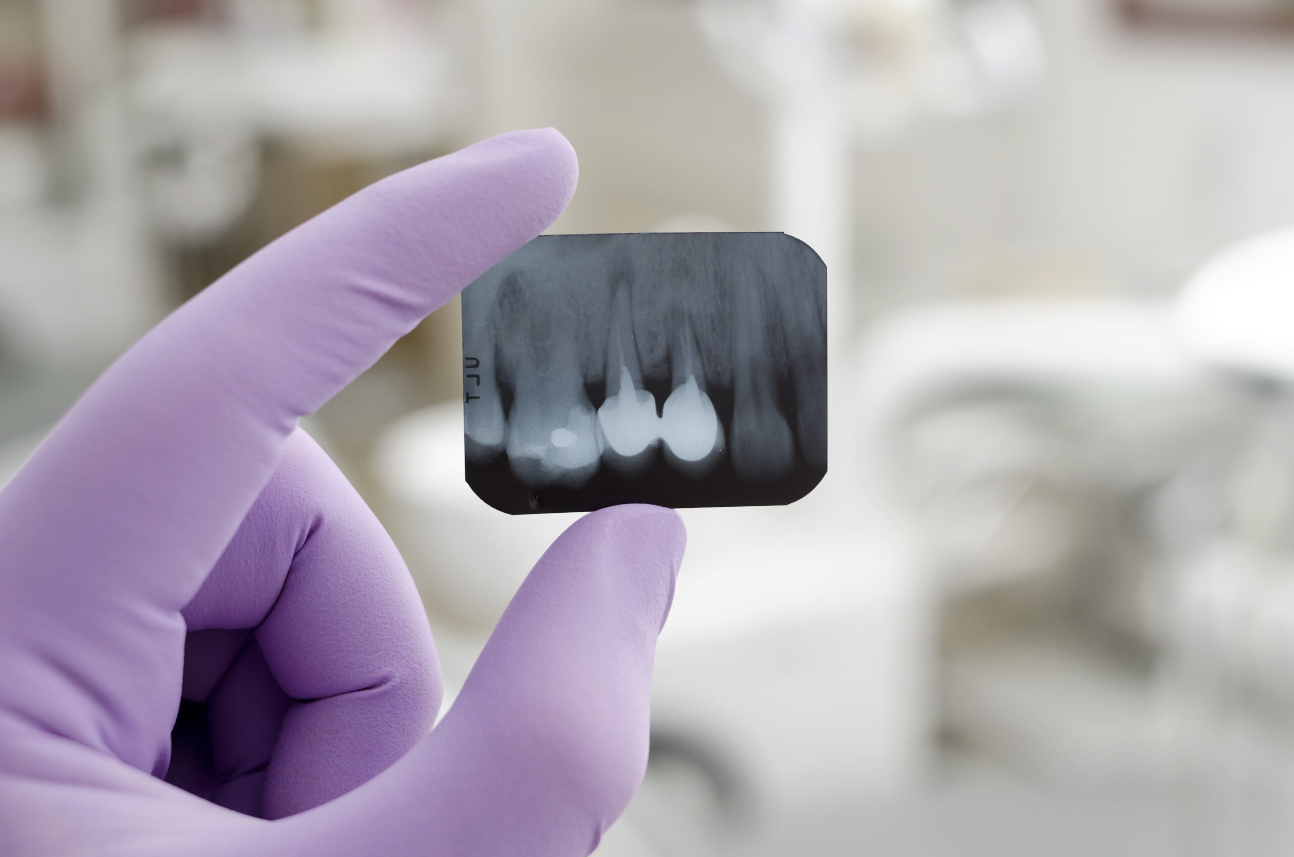

Painless and safe

Excellent detail

Digital access to diagnostics
What can a single-tooth x-ray reveal?
A periapical x-ray allows the detection of:
- Early-stage caries, including areas beneath existing fillings.
- Dental plaque deposits on smooth and damaged surfaces.
- Infection foci and inflammation zones.
- Position of unerupted or impacted teeth.
- Cysts, tumors, and other pathological formations.
- Cracks and chips in enamel and fillings, even in hard-to-reach areas of the oral cavity.
Thanks to advanced x-ray diagnostics technology, a single-tooth image provides critical information for planning treatments like root canal therapy, implantology, and other dental procedures.
Price: Single tooth X-ray
-
Dental point X-ray examination
50 PLN
How the Dynasty Stomatology dental clinic's loyalty program works
Learn more about the loyalty program
Step 01
Visit the clinic and scan the QR code at the reception desk
Step 02
Register online — enter your details
Step 03
Receive and accumulate points after each visit
Do you want to receive accurate modern diagnostics?
Schedule an appointment!
By clicking the button, you give your agreet your personal data in accordance with privacy policy.
Frequently asked questions about dental periapical X-rays
-
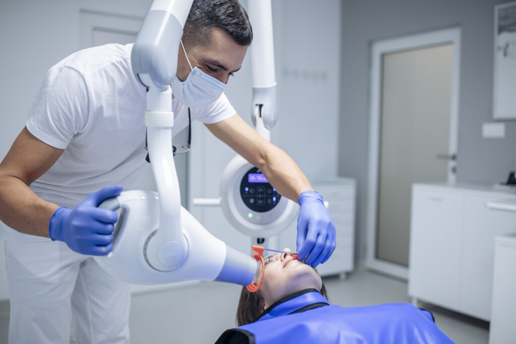
The patient is given a protective apron made of radiation-shielding material. The dentist places a special sensor either under the cheek or opposite the area being examined. Then the x-ray is taken and displayed on a computer monitor. The entire procedure lasts only a few minutes.
-
Dental x-rays help identify caries at various stages, pulpitis, periodontitis, pericoronitis, cysts, granulomas, and other abnormalities.
-
Yes, we recommend a targeted tooth x-ray both before treating dental pathologies and prior to implant procedures. This helps the doctor plan the treatment accurately and allows us to see the detailed condition of the teeth.
-
There are two main types of dental X-rays: intraoral and extraoral. Intraoral X-rays are taken inside the mouth and provide detailed images of individual teeth, helping us detect cavities, check tooth roots, and monitor bone health. Extraoral X-rays, such as panoramic images, are taken from outside the mouth and capture a broader view of the entire jaw, skull, and surrounding structures. Both types are safe, quick, and essential for accurate diagnosis and treatment planning.
-
A PA (periapical) X-ray captures the entire tooth from crown to root tip, including the surrounding bone — it's ideal for diagnosing infections, abscesses, or problems at the root level. Bitewing X-rays, on the other hand, show only the upper portions (crowns) of the upper and lower teeth together, making them perfect for detecting cavities between teeth and checking how well your fillings or crowns fit. In short, bitewings help us spot decay, while PAs give us the full picture of a tooth's root and the bone around it. Your dentist will choose the right type based on what needs to be examined.
-
A Pano (panoramic X-ray) is a single image that captures your entire mouth — all teeth, jaws, and surrounding structures — in one broad, sweeping shot. An FMX (Full Mouth X-ray series) is a set of 14–20 smaller, individual X-rays taken from different angles, providing much more detailed views of each tooth, its roots, and the surrounding bone. Think of a Pano as a wide landscape photo, while an FMX is like a collection of close-up portraits. Your dentist will recommend the right type based on whether they need a general overview or a detailed look at specific areas.
-
A target lesion on a dental radiograph appears as a small, well-defined dark (radiolucent) area surrounded by a ring of white (radiopaque) border, resembling a bullseye or target. It is most commonly associated with a condition called focal cemento-osseous dysplasia, which is a benign (non-cancerous) change in the bone around the tooth root. In most cases, this finding requires no treatment — just routine monitoring by your dentist to ensure it remains stable over time. If you notice this on your X-ray report, there's usually no reason to worry, but we're always happy to discuss it with you at your next visit.
Specialists who perform dental periapical X-rays
View all doctors
Anastasiia Danyliuk
General dentist, endodontist
Focuses not only on treatment effectiveness but also on patient comfort. Her main goal is to provide long-term results without the need for repeate...

Yulia Kryzhanovska
General dentist, endodontist
A doctor with a sense of humor who creates a positive atmosphere even in the dental chair.

Vladyslav Karkachov
General dentist, endodontist, prosthodontist
Relies on a well-thought-out treatment plan and good rapport with the patient. A friendly approach helps build trust, even among those afraid of de...

Yana Tereshchenko
General dentist, endodontist
Cares about both the health and the aesthetic and functional aspects of her patients’ teeth, helping preserve a natural, beautiful smile for years.

Yuliia Rudzynska
Pediatric dentist
Helps children not only maintain healthy teeth but also develop a positive attitude toward dentistry, ensuring fear-free visits.

Oleg Vus
General dentist, endodontist, prosthodontist
Oleg Vus is guided by the principles of professionalism, honesty, and patient care. He does not impose unnecessary procedures, but offers only the ...

Rostyslav Savruk
General dentist, endodontist, prosthodontist
Over 16 years of professional experience. Specialization in prosthetic dentistry and dental prosthetics. Use of advanced techniques and modern mate...

Borys Minakov
Prosthodontist
His approach focuses on individually tailored treatment plans that not only solve current issues but also prevent future ones. This helps patients ...

Dmytro Ostrovskyi
Prosthodontist
Over 30 hands-on courses completed in 9 years of practice. A comprehensive approach guarantees stable, durable, and predictable results.
Reviews of dental periapical X-rays in Krakow
Expert articles on the topic
View all blog-
25.08.2026
20
4 min. read
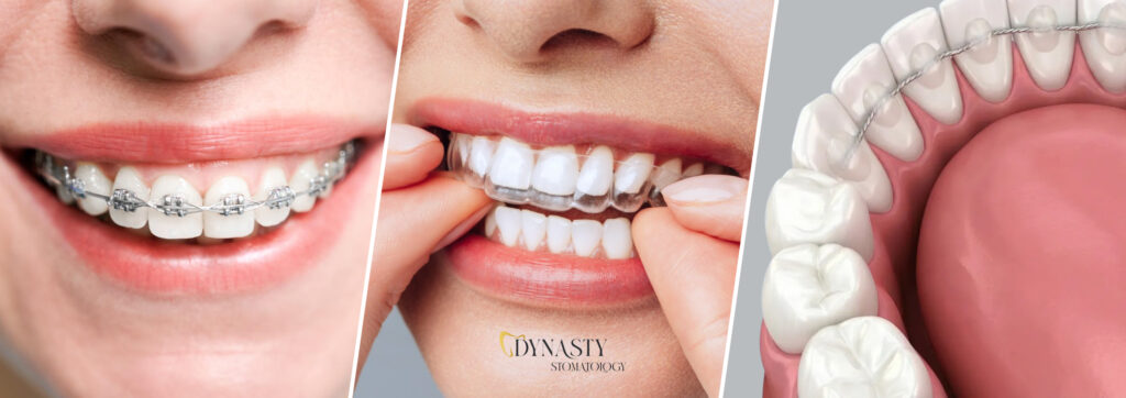
Braces, aligners or retainers – what to choose for bite alignment?
Malocclusion is not just about aesthetics. It can cause headaches, jaw overload, increased teeth clenching, speech problems, and even sleep…
Read more -
20.08.2026
8
11 min. read
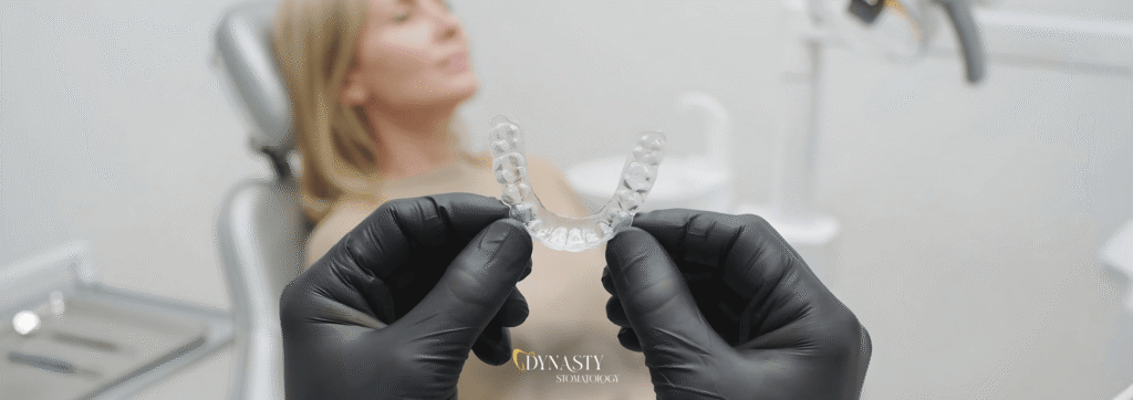
Retainer after removing orthodontic braces – what is it and what does it look like?
Retainer After Braces Removal – What Is It and What Does It Look Like? For patients who have completed treatment…
Read more -
07.08.2026
94
5 min. read
A new smile in 3 days — All-on-4 case in a complex clinical situation
Volodymyr came to Dynasty Stomatology with a clear request: to restore his teeth as quickly and efficiently as possible, improve…
Read more -
11.02.2026
485
8 min. read
What is Digital Dentistry — Everything you Need to Know
Until recently, a visit to the dentist was associated with significant discomfort. Uncomfortable impressions, long waits, and the need for…
Read more -
15.01.2026
539
8 min. read
Composite dental filling – what is it and when is it used?
For most people, a filling is a matter of appearance, comfort, and the assurance of long-lasting tooth health. It's important…
Read more -
12.12.2025
557
6 min. read
Can dental treatment be done during pregnancy – Dental care during pregnancy
Pregnancy is an important, but at the same time exciting and responsible event in a woman’s life. After all, in addition to being responsible for her own health, a woman is now responsible for the life of her child. During pregnancy, it’s important to monitor not only your general health but also your oral health. […]
Read more





Why is a single-tooth x-ray necessary?
A targeted dental x-ray image enables accurate diagnosis of various dental conditions and allows the dentist to prescribe effective treatment. Using a special digital device called radiovisiograph (RVG), we can capture detailed images of 1–3 adjacent teeth.
Advantages of a single-tooth x-ray.
Indications for a Diagnostic Tooth X-ray
During a dental examination, your dentist may suspect issues that cannot be accurately diagnosed without an x-ray image. A single-tooth x-ray is prescribed for well-founded medical reasons to identify such dental conditions as:
- Caries, both at early stages and underneath prosthetic crowns or fillings.
- Pulpitis.
- Periodontitis.
- Tooth fractures or dislocations.
- Endodontic treatment — to assess the condition of tooth roots and determine the number of canals.
- Chronic inflammation of the bone tissue surrounding the tooth.
- Cysts and granulomas.
- Evaluation of root canal filling quality during endodontic procedures.
- The condition of restored individual teeth after prosthetic treatments.
A tooth x-ray is also essential for determining the number, depth, and patency of root canals, ensuring treatment quality, and during prosthetic planning. It is also indicated in other clinical cases requiring a precise single tooth image.
Types of Dental X-ray Images
Modern digital x-ray technology allows us to obtain two main types of dental images:
-
Interproximal radiograph — displays and allows analysis of the anatomical condition of the crown part of a tooth, crucial for orthodontic treatment and dental care.
-
Periapical x-ray — focuses on the surrounding soft tissues of the tooth root, helping to detect infections, bone structures abnormalities, and to assess readiness for implant procedures.
In addition to intraoral single-tooth x-rays, comprehensive diagnostics may involve a panoramic x-ray (or pantomogram) to visualize the entire dentition, jaw structures, sinuses, and temporomandibular joints. For more complex cases, computed tomography (CT) is used to plan root canal treatment, tooth extraction, or implantology procedures.
At our x-ray lab, we use modern equipment that guarantees a high-quality, accurate image with minimal radiation exposure. This ensures the best possible diagnosis and treatment plan for each patient.
Price for a Single Tooth X-ray
Dynasty Stomatology, a modern dentistry center in Poland, is equipped with state-of-the-art x-ray machines and advanced diagnostic tools. Our digital radiovisiography system ensures maximum safety — it emits minimal radiation, making it suitable even for pregnant women. There are virtually no contraindications for this type of radiological diagnostics.
Our x-ray lab uses innovative technology that allows us to obtain high-resolution images of teeth, with precise scaling and zooming options. This guarantees an accurate image of individual teeth, their roots, and surrounding tissues. Despite offering premium-quality diagnostics, we maintain highly competitive pricing. The final cost depends on the type of point x-ray, its purpose, and the number of images required.
For more comprehensive diagnostics, we offer panoramic dental x-rays (also known as orthopantomograms) to assess the condition of the entire dentition, including upper and lower teeth, teeth and gums, and jaw structures. In cases that require even more detail, such as planned surgical treatments or implant procedures, we recommend computed tomography (CT), i.e., 3D diagnostic imaging.
At Dynasty Stomatology, we use only certified, modern equipment to ensure reliable diagnosis and treatment planning. Whether you need an image of a single tooth or a full pantomographic panoramic photo, our experienced medical staff will help you choose the right option for your dental treatment.
If you want to schedule an appointment or learn more about the procedure and diagnostics, feel free to contact us! Our office is equipped with modern, certified equipment that allows the doctor to assess the condition of your teeth and plan the treatment with a detailed treatment plan, ensuring the best care for you.
Schedule an appointment today for a consultation and initial examination. We are looking forward to seeing you!